Ultrasound: B-Scan
In an ultrasound B-scan outlines ofstructures are produced by using a multitransducer probe. Images of sections through the body are displayed on a screen. In a B-scan the echo signals are not applied to the Y-plates of the oscilloscope but are used instead to control the brightness of the spot on the oscilloscope screen.
Voltages from potentiometers which record the position and orientations of the transducer: are fed to the X- and Y-plates and determine the position of the spot on the screen. The co-ordinates must be accurate, and in a B-scan the error in the position of the spot on the screen is within 0.15 mm.
Rocking the probe
It should be noted that echoes are received only if the interface is normal to the beam of ultrasound. The interfaces inside the body are generally curved and at different orientations to each other; so, in order to obtain the best information about an interface, the probe may need to be rocked while it is moved on the surface of the body. This rocking is a skilful operation since the probe must be kept in contact with the surface; otherwise any air space will reflect the ultrasound before it can enter the body.
In order to ease the movement of the probe and to provide the required coupling between the transducer and the surface, oil is smeared on the patient's skin.
Uses of the B-Scan
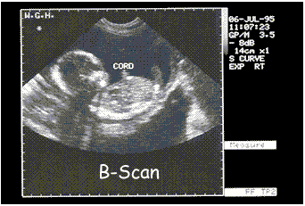
The largest diameter of the foetal head perpendicular to the midline is the bi-parietal diameter and is measured on the A-scan. In order to select the correct place for the A-scan, a B-scan is used to search for an echo from the midline structure of the brain. Such an echo produces a sharp line bisecting the image of the foetal skull.
 An important use of the B-scan in obstetrics is to determine the position of the placenta in relation to the entrance to the cervix. The condition called placenta praevia, ie when the placenta covers the entrance to the cervix, can then be diagnosed and the baby delivered by Caesarean section.
A further use of the B-scan is prior to and during amniocentesis, which involves inserting a tube into the uterus to remove some fluid and cells surrounding the foetus in order to carry out a genetic test on the chromosomes of the cells of the foetus. The position of the foetus and placenta have to be located accurately, which is done by the B-scan.