
 |
The Eye - Advanced level The eye is able to detect light energy - electromagnetic waves with wavelengths in the approximate range 380 nm to 760 nm. It therefore allows us to perceive objects which emit or reflect radiation in this range - radiation we call visible light. The eye has two physics systems within it:
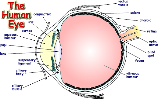
Eye Structure The diagram below has a few more items on it than the GCSE version. Well, you are doing A Level now! You need to be sure you know the parts in bold print and that you could label them on a diagram.
The eyeball is surrounded by a tough, fibrous coating, called the sclera, which is transparent at the front to form the cornea but white and opaque elsewhere. The choroid is tissue containing a network of blood vessels. These supply food and oxygen to the eye. It forms the lining between the sclera and retina. Melanin, a dark coloured pigment, helps the choroid limit reflection within the eye that could result in the perception of confusing images The thickened edge of the choroid in the region round the lens is called the ciliary body and contains circular muscle fibres, the ciliary muscles. These are responsible for altering the shape of the lens to give fine tuning of the focus of an image. These are involuntarily controlled by the brain when you focus on an object. Inside the choroid, at the back of the eye, is the retina, the light-sensitive layer, onto which light is focused by the lens. The fovea is at the centre of the retina. This tiny area is responsible for our central, sharpest vision. A healthy fovea is key for reading, watching television, driving, and other activities that require the ability to see detail. Unlike the peripheral retina, it has no blood vessels. Instead, it has a very high concentration of cones allowing us to appreciate fine detailed colour images. The lens is held in position by a circular band of tough, fibrous material called the suspensory ligament. The outer rim of this is attached to the ciliary muscle. Even when this muscle is relaxed there is a degree of tension in the ligament, keeping the lens slightly curved; in this position the lens has its maximum focal length. This will alow a normal eye to focus on objects at distances from 5 metres away to infinity, but a shortsighted eye will be unable to focus on distant objects even when the lens is at its least curved. Accomodation is the focussing process used to look at objects closer than five metres away. Images are brought into focus by contraction of the ciliary muscle, causing a reduction in tension in the lens and therefore an increase in its curvature and a consequent decrease in its length. The vid clip below explains this nicely!
The two chambers into which the eye is divided by the lens are filled with transparent fluid. The gelatinous vitreous humour is between lens and retina and the watery aqueous humour between the lens and the cornea. The pressure of these fluids on the sclera maintains the approximately spherical shape of the eye. A coloured extension of the ciliary body is known as the iris. The iris is a coloured ring of muscle with a variable aperture - the pupil. The aperture size is automatically controlled by the brain. It adjusts the iris size to allow sufficient light to enter it - enough to form an image. If too much light entered the eye the retina cells would 'max out' and the detail would be indestinguishable, therefore in bright light the aperture of the iris (the pupil) is very small. In low light conditions the aperture is made as wide as possible to allow images of dim objects to be perceived.
The diameter of the pupil also decreases when close up objects are viewed. This has the effect of removing the outermost rays of light which may not be refracted sufficiently to obtain a sharp image; hence the size of the pupil is used to control spherical aberration. Click here to go to a page on:
There are a set of A level standard questions on this topic. Here is the link. You may wish to look at the other topics that are covered at A level standard. If so use the upper left hand menu (pale blue and select practice questions to navigate to the menu page. |
Follow me...
|


Cyberphysics - a web-based teaching aid - for students of physics, their teachers and parents....