A level: Ultrasound Questions
Q6.The diagram shows a cross-section through an ultrasound generator.
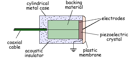
(a) Explain the purpose of the backing material.
It damps the oscillation of the crystal to zero quickly  when the driving signal is removed
(2 marks)
(b) State the main difference between the probe used in an A-scan and that used in a B-scan.
An A-scan has a single transducer, but a B-scan uses a multi-transducer probe
(1 mark)
(c) The diagram belowshows the display on an oscilloscope screen of an A-scan to find the size of an organ. The speed of sound in the organ is 1200 m s-1
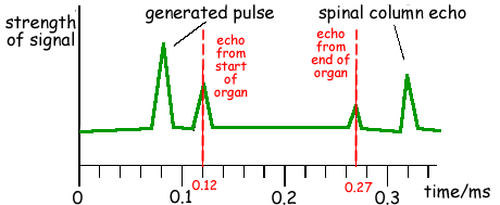
(i) Calculate the diameter of the organ.
Time for pulse to go the double distance
= 0.27 – 0.12 = 0.15 ms
diameter = (1200 × 0.15 × 10-3)/2 = 0.090 m
(ii) State two processes which reduce the strength of the reflected signal received by the probe.
You only get a partial reflection at boundary You also get attenuation as the signal passes through body
(4 marks)
(Total 7 marks)