'A' Level Medical Option Questions - The Heart - ECG
Q3. The graph below shows the variation of membrane potential difference with time of a nerve fibre, known as an action potential.
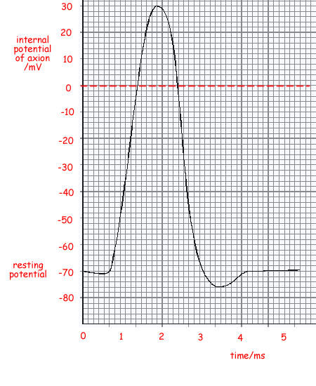
(a) Complete the graph by adding suitable axes, units and scales.
- axes: time/ms, action potential/mV

- time scale can range about 5 ms - usually about 2-3 ms for central peak
- action potential scale +30 mV to –70 mV
3 marks
(b) Describe the processes involved in the production of such an action potential when a nerve is stimulated.
- Na+ ions move into the cell .
- The pd rises (from –70 to 0) , this is called depolarisation
- K+ ions move out of the nerve . The pd returns/falls to –70/resting potential, this is called repolarisation
- Na+ moving from 0 to +30 called reverse polarisation
- To restore starting equilibrium of ions, the Na/K pump operates
3 marks
Total 6 marks