'A' Level Medical Option Questions - X-Rays
Q1. The simplified diagram shows a modern X-ray tube.
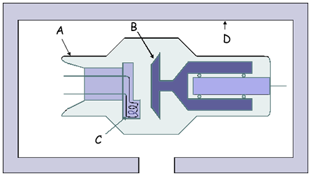
(a) For each of the labelled parts, state what it is and explain its purpose.
(8 mark)
(b) On the diagram draw and label
(i) the direction of the electron beam,
(ii) the direction of the useful X-ray beam.
(2 marks)
(Total 10 marks)

Q2.
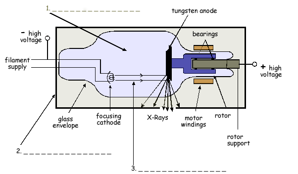
(a) The diagram shows a rotating-anode X-ray tube. Complete the labelling of the three numbered arrows in the diagram.
(3 marks)
(b) Explain why the anode
(i) is rotated,
(ii) has a bevelled edge.
(3 marks)
(c) Define for a material,
(i) the linear attenuation coefficient, 
(ii) the half-value thickness.
(2 marks)
(d) A monochromatic X-ray beam of intensity 6.0 W m–2 is incident on an aluminium sheet of thickness 2.0 mm. For these X-rays, the half-value thickness of aluminium is 3.2 mm. Calculate the intensity of the transmitted beam.
(3 marks)
(Total 11 marks)
Q3.
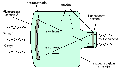
The diagram shows a fluoroscopic image intensifier.
(a) State the purpose of:
(i) the fluorescent screen, A,
(ii) the photocathode,
(iii) the anodes,
(iv) the fluorescent screen, B.
(4 marks)
(b) Give one example of a medical application for which an image intensifier might be used. Explain why the use of an image intensifier is required.
(2 marks)
(Total 6 marks)
Q4. Diagnostic X-rays are produced using a rotating anode X-ray tube.
(a)
(i) State two methods which can be used to increase the intensity of the X-ray beam produced by the tube.
(ii) For each method of increasing intensity, state the effect on the maximum X-ray photon energy.
(3 marks)
(b) Before taking an X-ray photograph, the X-ray beam emerging from the tube is passed through an aluminium filter. State and explain the reason for filtering the X-rays.
(3 marks)
(Total 6 marks)
Q5.
(a) When an X-ray image is obtained of certain organs, image contrast enhancement is necessary. Explain why image contrast enhancement is needed and describe how this might be achieved.
(3 marks)
(b) A monochromatic X-ray beam of intensity 3.2 × 10⁻² W m–2 is incident on an aluminium sheet. Calculate the thickness of aluminium required to reduce the intensity of the X-ray beam to 1.2 × 10⁻² W m–2.
mass attenuation coefficient of aluminium, m = 0.012 m2 kg–1
density of aluminium,  = 2700 kg m–3
(3 marks)
(Total 6 marks)
Q6. In the course of diagnosis and treatment of a child's broken arm, several images of the arm are required. Similarly, to check the progress of a woman's pregnancy, several images of the foetus are required.
In each case, state which imaging technique would probably be used and give two reasons for the choice.
(a) Broken arm
(b) Foetus
(Total 4 marks)
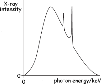
Q7.
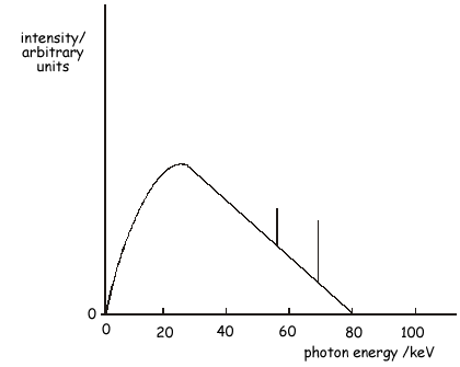
(a) An X-ray tube operates with a pd across the tube of 80 kV. The figure above shows the X-ray spectrum emitted. Explain why the spectrum has spikes at specific photon energies.
(2 marks)
(b) The pd across the tube is increased to 90 kV. Sketch on the figure above the X-ray spectrum produced at this new pd.
(3 marks)
(c) At the working pd of 80 kV, the anode current was 120 mA. The X-ray tube has an efficiency of 0.70 %. Calculate the rate of production of heat at the anode.
(3 marks)
(Total 8 marks)
Q8.
The figure below shows the design of an X-ray image intensifier. The main components are labelled A to D.
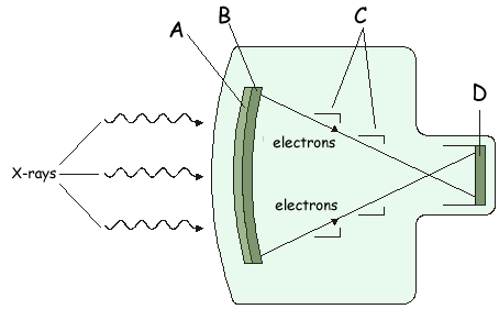
Name each component and state its purpose in the process of image intensification.
(Total 8 marks)
Q9.
(a) Explain what is meant by the half-value thickness of lead for X-rays.
(b) Calculate the linear attenuation coefficient of lead for 90 keV X-ray photons. half value thickness of lead for 90 keV X-ray photons = 12mm.
(c) Calculate the thickness of lead needed to reduce the intensity of a beam of 90 keV X-ray photons to 5.0% of the intensity incident on the lead.
(Total 6 marks)
Q10.
(a) The X-ray spectrum for a certain X-ray tube target is shown below. Explain the process which gives rise to spikes at certain photon energies.
(3 marks)
(b) A film cassette, placed under a patient being X-rayed, is shown below:
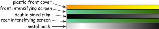
Explain how the intensifying screens in the film cassette achieve their purpose and state
their benefit to the patient.
(3 marks)
(Total 6 marks)
Q11.
(a) In an X-ray tube, electrons are accelerated from rest through a pd of 72.4 kV before they hit the target anode.
(i) Calculate the kinetic energy of an electron as it reaches the anode. Give your answer to an appropriate number of significant figures.
(2 marks)
(ii) Assuming that the electron gives up all this energy to form an X-ray photon, calculate the wavelength of the photon.
(b) X-rays are used in a CT scanner. Describe briefly how a CT scanner produces an image.
(3 marks)
(7 marks total)
Q12.
(a) Explain the term contrast enhancement when applied to X-ray photographic imaging and explain how such enhancement is achieved.
(2 marks)
(b) The half-value thickness of lead for 90 keV X-ray photons is 12 mm.
(i) What is meant by half-value thickness?
(ii) Calculate the linear attenuation coefficient, μ, for these X-ray photons in lead.
(iii) It is required to reduce the intensity of X-radiation escaping in unwanted directions from a 90 kV X-ray tube to 5% of its full intensity. Calculate the thickness of lead shielding needed to achieve this reduction.
(5 marks)
(7 marks total)