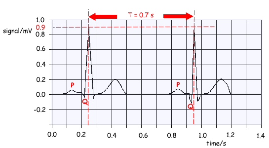
'A' Level Medical Option Questions - The Heart - ECG
Q1.
The graph below shows a normal electrocardiogram (ECG) signal obtained at the surface of the skin of a patient.
(a) What is the amplitude of the main pulse in the signal?
Pulse amplitude: 0.9 mV 
1 mark
(b) Find the period of the heart beat and from it calculate the pulse rate per minute.
T = 0.7s 
f = 1/T = 1/0.7 Hz
60/.07 = 86 min–1 
2 marks
(c) What changes would you expect to see in the electrocardiogram if the patient began to take exercise?
T would get shorter
as the flat part of trace would shrink 
2 marks
(d) On the graph, label with a P the points where atrial contraction occurs and with a Q the points where ventricular contraction starts.
See trace above for P and Q location 
2 marks
Total 7 marks