 |
||||
    |
||||
 Positron
Emission Tomography Positron
Emission Tomography |
||||
|
Positron
emission tomography, also called PET imaging or a PET scan, is a diagnostic
examination that involves the building up of 3-d images by detecting
the concentration of radiation emission at various The tracer used is a positron emitter. Positrons are anti-matter beta-particles emitted from the radioactive substance administered to the patient. When a positron interacts with an electron annihilation occurs and two gamma rays are produced that fly off in opposite directions. The PET scanner detects this pair of gamma rays making it possible to compute where they originated from in the body - this will be the point where the positron was emitted, as a positron won't get far in matter after emission. So... the PET scanner detects gamma rays - but the tracer emits positrons!
PETscanning is non-invasive but does involve ionizing radiation.The images of the human body developed with this technique can be used to evaluate a variety of diseases. Common uses of the procedure
The equipment
The ProcedureBefore the examination begins, a positron-emitting radioactive substance is produced in a machine called a cyclotron and attached, or tagged, to a natural body compound, most commonly glucose, but sometimes water or ammonia. Oxygen 15 is a typical radioisotope used in PET scanning, it decays with a half-life of 2.25 minutes by the emission of positrons. The isotope is prepared by irradiation of nitrogen by means of cyclotron-accelerated deuterons. The cyclotron needed for this procedure is small enough power to be on site in a large hospital complex (although it is an expensive piece of equipment and needs expert operation!). The doctor is interested in finding out where this natural body compound accumulates. From that information s/he can wok out how well body organs are functioning. Once this tagged substance is administered to the patient, the radioactivity localizes in the appropriate areas of the body and can then be detected by the PET scanner.Therefore the doctor gains information on the use of that compound by the body.
How the procedure is performedA nurse or technologist takes the patient into a special injection room, where the radioactive substance is administered as an intravenous injection (although in some cases, it will be given through an existing intravenous line or inhaled as a gas). It then takes approximately 30 to 90 minutes for the substance to travel through the body and accumulate in the tissue under study. During this time, the patient will be asked to rest quietly and avoid significant movement or talking, which may alter the localization of the administered substance. After that time, scanning begins. This may take 30 to 45 minutes. Some patients, specifically those with heart disease, may undergo a stress test in which PET scans are obtained while they are at rest and again after undergoing the administration of a pharmaceutical to alter the blood flow to the heart. Usually, there are no restrictions on daily routine after the test, although the patient should drink plenty of fluids to flush the radioactive substance from his/her body as quickly as possible after the procedure and for a limited time the patient might be asked to avoid public transport or places where there could be a pregnant woman or a young child - as they will be a source of gamma rays for a while - until the emission drops to background level.This will depend on the tracer used and its half life. The shorter the half life the quicker the level will drop to 'normal'. Oxygen tagged procedures are very quick to become benign because the half life is so short. What do you experience during the procedure?
You will not feel anything
related to the radioactivity of the substance in your body. |
||||
 |
 |
|||
 points
within the body. This means that a picture of where the radioactive tracer
has accumulated allows doctors to understand how the body is functioning
and they can use that information to decide upon what treatment is suitable.
points
within the body. This means that a picture of where the radioactive tracer
has accumulated allows doctors to understand how the body is functioning
and they can use that information to decide upon what treatment is suitable.
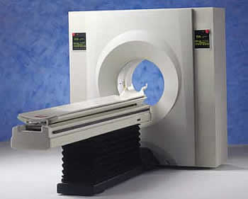 A PET scanner looks
like a large doughnut - it has a hole in the middle. Within this machine
are multiple rings of detectors that record the gamma rays emitted when
a positron from the radioactive substance in your body annihilates an
electron in the patient's body. It only 'counts' gamma rays pairs. The
information collected permits a 3-d image of the body to be obtained.
While lying on a cushioned examination table, the patient is moved into
the hole of the machine. The images are displayed on the monitor of a
nearby computer, which is similar in appearance to a home personal computer.
A PET scanner looks
like a large doughnut - it has a hole in the middle. Within this machine
are multiple rings of detectors that record the gamma rays emitted when
a positron from the radioactive substance in your body annihilates an
electron in the patient's body. It only 'counts' gamma rays pairs. The
information collected permits a 3-d image of the body to be obtained.
While lying on a cushioned examination table, the patient is moved into
the hole of the machine. The images are displayed on the monitor of a
nearby computer, which is similar in appearance to a home personal computer.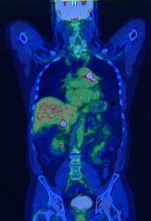 The
data from the PET scanner can be coded into colour computer images. Different
colours or degrees of brightness on a PET image are made to represent
different levels of tissue or organ function because they represent the
concentration of annihilation events at that location. Because healthy
tissue uses glucose for energy, it accumulates some of the tagged glucose,
which will show up on the PET images. However, cancerous tissue, which
uses more glucose than normal tissue, will accumulate more of the substance
and appear brighter than normal tissue on the PET images.PET can shows
blood flow in the brain by imaging trace amounts of radioisotopes at various
locations. If it indicates a decrease in blood flow to parts of the brain
this can indicate such diseases as Alzheimer's.
The
data from the PET scanner can be coded into colour computer images. Different
colours or degrees of brightness on a PET image are made to represent
different levels of tissue or organ function because they represent the
concentration of annihilation events at that location. Because healthy
tissue uses glucose for energy, it accumulates some of the tagged glucose,
which will show up on the PET images. However, cancerous tissue, which
uses more glucose than normal tissue, will accumulate more of the substance
and appear brighter than normal tissue on the PET images.PET can shows
blood flow in the brain by imaging trace amounts of radioisotopes at various
locations. If it indicates a decrease in blood flow to parts of the brain
this can indicate such diseases as Alzheimer's.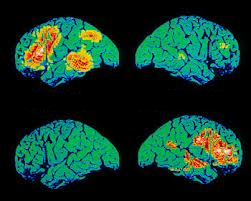 The
administration of the radioactive substance feels like a slight pinprick
if given by intravenous injection. You will then be made as comfortable
as possible before you are positioned in the PET scanner for the test.
You will be asked to remain still for the duration of the examination.
Patients who are claustrophobic may feel some anxiety while positioned
in the scanner. Also, some patients find it uncomfortable to hold one
position for more than a few minutes.
The
administration of the radioactive substance feels like a slight pinprick
if given by intravenous injection. You will then be made as comfortable
as possible before you are positioned in the PET scanner for the test.
You will be asked to remain still for the duration of the examination.
Patients who are claustrophobic may feel some anxiety while positioned
in the scanner. Also, some patients find it uncomfortable to hold one
position for more than a few minutes. ![]()