|
See
also loudness perception and dBA/dB
scales
The
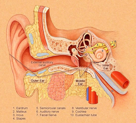
above image was taken from http://www.harford.cc.md.us/Faculty/wrounds/ear.gif
The ear
has three main parts:
The outer
ear (the part you can see) opens into the ear canal. The eardrum separates
the ear canal from the middle ear. Small bones in the middle ear
help transfer sound to the inner ear. The inner ear contains the
auditory (hearing) nerve, which leads to the brain.
Any source of sound
sends vibrations or sound waves into the air. These funnel through the
ear opening, down the ear, canal, and strike your eardrum, causing it
to vibrate. The vibrations are passed to the small bones of the middle
ear, which transmit them to the hearing nerve in the inner ear. Here,
the vibrations become nerve impulses and go directly to the brain, which
interprets the impulses as sound.
Click here
for a page to take you through revision of how sound is propagated.

A
collective name for the bones in the ear is the ossicles
A
collective name for the vestibular and cochlear nerves is the auditory
nerve
Check
your knowledge of this by trying the crossword
 The pinna,
the outer part of the ear, serves to "catch" the sound waves.
It also helps you determine the direction of a sound. Your brain determines
the horizontal position of a sound by comparing the information coming
from your two ears. The pinna,
the outer part of the ear, serves to "catch" the sound waves.
It also helps you determine the direction of a sound. Your brain determines
the horizontal position of a sound by comparing the information coming
from your two ears.
Once
the sound waves travel into the ear canal, they vibrate the tympanic
membrane or eardrum. This is a thin, cone-shaped piece of skin,
about 10 millimeters (0.4 inches) wide. It is positioned between the ear
canal and the middle ear. Air from the atmosphere flows in from your outer
ear onto one side and from your mouth on the other (the middle ear is
connected to the throat via the eustachian tube) so the air pressure on
both sides of the eardrum remains equal. This pressure balance lets your
eardrum move freely back and forth with even the slightest air-pressure
fluctuations. Higher-pitch sound waves move the drum more rapidly (higher
frequency), and louder sound moves the drum a greater distance
(bigger amplitude). The eardrum is the entire sensory element
in your ear. The rest of the ear serves only to pass along the information
gathered at the eardrum.
For the
most part, the changes in air pressure due to sound waves we hear are
extremely small. They don't apply much force on the eardrum, but the eardrum
is so sensitive that this minimal force moves it a good distance. However,
the cochlea in the inner ear conducts sound through a fluid, instead of
through air. This fluid has a much higher inertia than air so the small
force felt at the eardrum is not strong enough to move this fluid. Therefore
before the sound passes on to the inner ear, the total pressure (force
per unit of volume) must be amplified.
This amplification
is caried out by the ossicles:
- The malleus
- the hammer
- The incus - the anvil
- The stapes - the stirrup
The malleus
is connected to the center of the eardrum, on the inner side. When the
eardrum vibrates, it moves the malleus from side to side like a lever.
The other end of the malleus is connected to the incus, which is
attached to the stapes. The other end of the stapes rests against
the cochlea, through the oval window.
When air-pressure
compression pushes in on the eardrum, the ossicles move so that
the stapes pushes in on the cochlear fluid. When air-pressure rarefaction
pulls out on the eardrum, the ossicles move so that the stapes pulls in
on the fluid. Essentially, the stapes acts as a piston, creating
waves in the inner-ear fluid to represent the air-pressure fluctuations
of the sound wave.
The ossicles amplify
the force from the eardrum in two ways:
- The main amplification
comes from the size difference between the eardrum and the stirrup.
The eardrum has a surface area of approximately 55 square millimeters,
while the faceplate of the stapes has a surface area of about 3.2 square
millimeters. Sound waves apply force to every square inch of the eardrum,
and the eardrum transfers all this energy to the stapes. When you concentrate
this energy over a smaller surface area, the pressure (force per unit
of volume) is much greater.
- The configuration
of ossicles provides additional amplification. The malleus is longer
than the incus, forming a basic lever between the eardrum and
the stapes. The malleus moves a greater distance, and the incus moves
with greater force (energy = force x distance).
This amplification
system is extremely effective. The pressure applied to the cochlear fluid
is about 22 times the pressure felt at the eardrum. This pressure amplification
is enough to pass the sound information on to the inner ear, where it
is translated into nerve impulses the brain can understand by the cochlea.
The cochlea structure
consists of three adjacent tubes separated from each other by sensitive
membranes. These tubes are coiled in the shape of a snail shell. The membrane
between these tubes is so thin that sound waves travel as if the tubes
weren't separated at all. The stapes moves back and forth, creating pressure
waves in the entire cochlea. The round window membrane separating
the cochlea from the middle ear gives the fluid somewhere to go. It moves
out when the stapes pushes in and moves in when the stapes pulls out.
Click here
for a diagram of how this works (it 'opens out' the snail shell arrangement
for clarification!)
The middle membrane,
the basilar membrane, is a rigid surface that extends across the length
of the cochlea. When the stapes moves in and out, it pushes and pulls
on the part of the basilar membrane just below the oval window. This force
starts a wave moving along the surface of the membrane. The wave travels
something like ripples along the surface of a pond, moving from the oval
window down to the other end of the cochlea.
The basilar membrane
has a peculiar structure. It's made of 20,000 to 30,000 reed-like fibers
that extend across the width of the cochlea. Near the oval window, the
fibers are short and stiff. As you move toward the other end of the tubes,
the fibers get longer and less rigid. This gives the fibers different
resonant frequencies. A specific wave frequency will resonate perfectly
with the fibers at a certain point, causing them to vibrate with a big
amplitude. Because of the increasing length and decreasing rigidity of
the fibers, higher-frequency waves vibrate the fibers closer to the
oval window, and lower frequency waves vibrate the fibers at the
other end of the membrane.
The organ of corti
is a structure containing thousands of tiny hair cells. It lies on
the surface of the basilar membrane and extends across the length of the
cochlea.
Until a wave reaches
the fibers with a resonant frequency, it doesn't move the basilar
membrane very much at all. But when the wave finally does reach the resonant
point, the membrane suddenly releases a burst of energy in that area.
This energy is strong enough to move the organ of corti hair cells at
that point.
When these hair
cells are moved, they send an electrical impulse through the
cochlear nerve. The cochlear nerve sends these impulses on to the
cerebral cortex, where the brain interprets them. The brain determines
the pitch of the sound based on the position of the cells sending electrical
impulses. Louder sounds release more energy at the resonant point along
the membrane and so move a greater number of hair cells in that area.
The brain knows a sound is louder because more hair cells are activated
in an area.
|


