Applications of Ultrasound
Examples of various probe design related to probe use:
A trans-oesophageal probe is shown in the diagram below. So that the transducer array is nearer to
the patient's heart
the narrow flexible tube is swallowed by the patient. This avoids
having to
scan between the gaps in a patients ribs.

|
 In an obstetric scan the probe used is usually one that looks like a curved soap bar (more accurately known as a 'convex-array' transducer, and the one with a flat surface a 'linear-array' transducer ) which can be slid over the maternal abdomen while maintaining good surface contact to the abdominal surface for the whole width of the probe. It has no moving parts. It is comparatively inexpensive and can be used by relatively unskilled operators. In an obstetric scan the probe used is usually one that looks like a curved soap bar (more accurately known as a 'convex-array' transducer, and the one with a flat surface a 'linear-array' transducer ) which can be slid over the maternal abdomen while maintaining good surface contact to the abdominal surface for the whole width of the probe. It has no moving parts. It is comparatively inexpensive and can be used by relatively unskilled operators.
A frequency of between 1-3 MHz is suitable for abdominal imaging as low frequency waves produce low resolution imaging and increasing attenuation occurs at very high frequency.
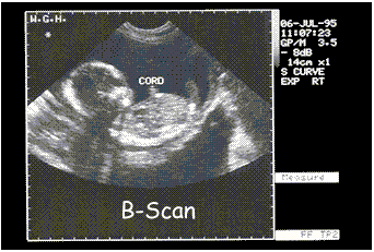
Pregnancy problems can be detected and general progress quantitatively monitored by using A-scans for accurate measurements and B-scans for general development.
 A B-Scan produces a sectional 2-D image. Each point on the monitor represents echo amplitude by 'grey-scale' representation. The brighter the point on the screen the 'louder' the echo. This is used to identify the part of the foetus to be measured. A multiple array of transducers is used to gain the information that builds up into the 2-d representation, whereas an A-scan probe only needs a single transducer. A B-Scan produces a sectional 2-D image. Each point on the monitor represents echo amplitude by 'grey-scale' representation. The brighter the point on the screen the 'louder' the echo. This is used to identify the part of the foetus to be measured. A multiple array of transducers is used to gain the information that builds up into the 2-d representation, whereas an A-scan probe only needs a single transducer.
|
In a vaginal scan, the probe
has to be a long and slender piece to fit into the vagina.

|
 Accurate distances within the eye can be measured using a type of probe designed especially for ophthalmic use. It can be used to measure the thickness of the cornea and be used to ascertain whether a person is suitable for laser eye surgury. Accurate distances within the eye can be measured using a type of probe designed especially for ophthalmic use. It can be used to measure the thickness of the cornea and be used to ascertain whether a person is suitable for laser eye surgury.
The scanner is either placed in direct contact with the eye or via a water bath (less risk of damage to eye surface) it therefore needs to be small and the transducer head suited to the curvature of the eye. It is used in A-scan mode with a frequency of 8-13MHz. 
- a = cornea spike
- b = anterior lens spike
- c = posterior lens spike
- d = retinal spike
- e = orbital spike
|
 The anatomical structure within the kidney can be viewed using a common curvilinear probe with a frequency of 5MHz in B-scan mode. The anatomical structure within the kidney can be viewed using a common curvilinear probe with a frequency of 5MHz in B-scan mode.
The higher the frequency the better the resolution of the image - 5 MHz will give detail of structure to within 1mm. |
 The function of the heart valve is best viewed using a trans-oesophageal probe with a frequency of 2.0-5.0 MHz. M-Scan complemented by a B-scan. Without this specialized probe a simple sector scan or compound scan between the ribs would be necessary to avoid interference of them. Doppler imaging would allow blood flow into and out of the valve to be monitored and efficiency and any leakage can be assessed. The function of the heart valve is best viewed using a trans-oesophageal probe with a frequency of 2.0-5.0 MHz. M-Scan complemented by a B-scan. Without this specialized probe a simple sector scan or compound scan between the ribs would be necessary to avoid interference of them. Doppler imaging would allow blood flow into and out of the valve to be monitored and efficiency and any leakage can be assessed.
|


