
 |
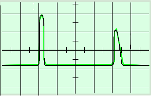
Ultrasound: A-scan The A-scan system is used where precise locations of internal boundaries are to be measured. To perform an A-Scan you only need a probe with a single transducer. An A scan relates the amplitude of a signal with the strength of reflection. To prepare the patient for a scan a coupling gel is smeared onto the body to expel the air from between the probe and the patient's skin. The ultrasound transducer or probe is set to emit short, sharp pulses of ultrasound of a few microseconds duration and linked to output data to a cathode ray oscilloscope. The probe head is then placed on the body of the patient.. At the same time as a pulse of ultrasound is emitted by the transducer, the spot on a cathode ray oscilloscope screen is triggered and starts moving across the screen. The ultrasound pulse travels into the body and, when it reaches an interface or boundary between different tissues in the body, some of the sound energy is reflected back towards the transducer where it causes an electrical signal to be generated. (For detail on how reflection occurs at tissue boundaries click here) The signal causes the spot on the screen to be deflected vertically for the duration of the received echo pulse. The spot then returns to its original level and continues to move across the screen. The returning echo pulse is also displayed as a deflection of the trace on the screen of the oscilloscope.
Since the pulse was emitted at the same time as the spot started to move across the screen, the displacement of the echo from the starting point is a measure of the time taken for the transmitted pulse to reach the first surface plus the time for the echo to return to the transducer. (2t) From the time-base setting of the oscilloscope and the screen visual display the operator can calculate the time taken for the ultrasound wave to move across an organ's depth = 1/2 t. The speed of ultrasound within a particular organ is known. Let us call this 'c'. The section depth 'd' of the organ can then be calculated using a simple calculation: d = 1/2 ct See practice questions for the type of calculation you will be asked to do. When a pulse of ultrasound passes through the body, the amplitude of the pulse is attenuated (some energy is absorbed by tissue - some passes through to other lays, some is scattered in directions that are not picked up by the probe). This means that echoes from surfaces further away from the transducer require to be amplified more than those from surfaces nearer the transducer. This amplification is achieved by a swept-gain amplifier linked to the time base of the oscilloscope.
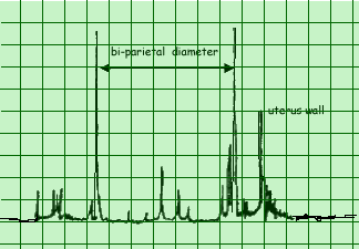
The A-scan method is used to measure the bi-parietal diameter (size of the foetal head) which may be used in order to determine the length of gestation and also to monitor the growth of the foetus. The greatest density difference is between the bone and tissue - therefore the two large spikes correspond to the beam being reflected from the near and far side of the baby's skull. To ensure the scan is taken from the correct angle a B-Scan is used. The A-scan technique is fine if the surfaces of interest within the body lie along a particular line. For more complex structures, however, it becomes very difficult to identify the surfaces which give rise to particular echoes, it is then useful to perform a B-Scan.
|
Follow me...
|


Cyberphysics - a web-based teaching aid - for students of physics, their teachers and parents....