
 |
The EYE: Photosensitive cells
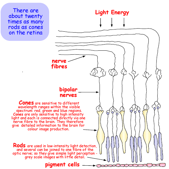
Nerve fibres lie on the surface of the retina. Light must penetrate this layer before activating the photosensitive cells beneath.The photosensitive cells are packed very closely together and are light-sensitive along their length; therefore the eye can focus simultaneously on objects between infinity and about five metres, without adjustment of the lens (without accommodation). There are two types of photosensitive cells - rods and cones. The photons absorbed cause light-sensitive chemicals in the those cells to decompose and this stimulates electrical impulses in the associated nerve fibres leading to the brain. The size of the voltage produced indicates the intensity of light falling on them (the number of photos per second). The electrical signals produced are passed along the optic nerve, through to the visual cortex of the brain where the information is assimilated into visual information. The left hand visual cortex processes data from the left hand side of both the left and right eyes and the right hand visual cortex processes data from the right hand side of both the left and right eyes . There are two classes of photosensitive cells:
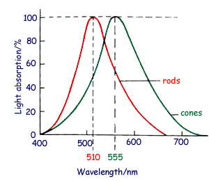
They contain the light-sensitive pigment rhodopsin. On exposure to light, rhodopsin is bleached, first yellow then clear, thereby stimulating the rod cell and its associated nerve cells. Bleached rhodopsin is rapidly restored to its original condition by an enzymatic process requiring vitamin A derived from the blood supply in the choroid. Although the rods do not supply any information concerning colour, they are not equally sensitive to all wavelengths, displaying maximum absorption in the green (500-580 nm) region of the spectrum, at about 510nm. Cones (for high detail, colour vision)
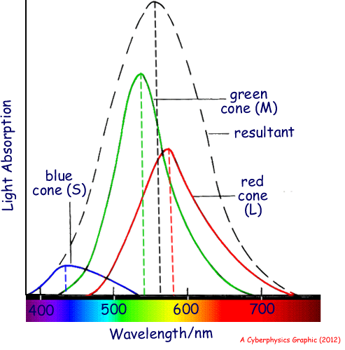
Cones are responsible for the more acute vision experienced in ordinary daylight conditions (colour or photopic vision). They contain light-sensitive proteins - photopsins and a retinene and, as in the rods, a bleaching process initiates the nerve impulse. There are three types of cone in the retina: Each cone type detects a different range of wavelength. Between them, they discern all wavelengths from about 380 nm to 760 nm, producing the greatest sensitivity at around 555 nm. The graph below shows the approximate contribution to overall cone absorption from the three groups, and illustrates why greens and yellows are normally perceived to be brighter colours than deep blues or reds.
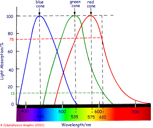
For example, monochromatic light of wavelength 610 nm stimulates the red cones to a 'stimulus value' of 75%, the green cones to 13% and the blue cones to 0%. The brain then interprets the stimulus ratios 75:13:0 as an orangy red, You can use the graph to determine what response each part of the spectrum gives - monochromatic stimulation.
|
Follow me...
|
 Rods (for black and white - monochrome vision)
 Rods are responsible for vision in dim light (night, twilight or
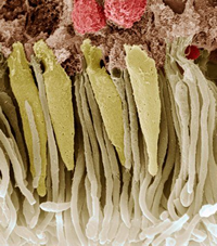


Cyberphysics - a web-based teaching aid - for students of physics, their teachers and parents....