A level: Ultrasound Questions
Q3. An ultrasound transducer is used to obtain an A-scan of an internal organ. The A-scan pulses shown on the diagram of the oscilloscope screen were identified as coming from the front and rear surfaces of the organ. The timebase setting was 0.02s/div.
(a) Describe the practical process, including details of the use of the transducer and the adjustment of the oscilloscope, required to produce this A-scan.
The surface of the body is covered with an acoustically matched gel (gel with similar density to human tissue) to improve transmission from the ultrasound transducer to body.  Short ultrasound pulses are sent into the body and the echoes received from boundaries between media of different densities are detected by the transducer. An oscilloscope is set up so that the sweep time is synchronised with the ultrasound pulse frequency
3 marks
(b) From the A-scan, estimate:
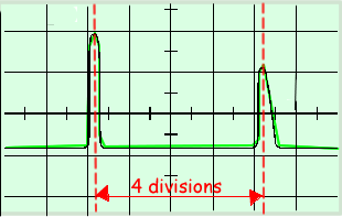
(i) the thickness of the organ if the speed of ultrasound in the tissue is 1500 ms–1 (the horizontal scale is 0.02 ms/cm),
thickness = 1/2 c  t
t
t = 4 x 0.02 = 0.08s (see diagram)
= 1/2 × 1500 × 0.08 × 10–3 (m)
= 0.06 m
(ii) the duration of the first ultrasound pulse.
pulse duration = 0.3 × 0.02 = 0.006ms
3 marks
(c) Give two reasons why the height of the second pulse is smaller than that of the first pulse.
Two from:
- Extra distance in tissue results in more signal absorption
- Smaller fraction of signal reflected at second surface
- The pulse will be more spread over time
- The signal is diffracted
2 marks
Total 8 marks